Click Here for More Images from iStock- 15% off with coupon 15FREEIMAGES 
Cross-section view of Mitochondria, 3d rendering. Computer digital drawing.
plant Flower bud of Lilium brownii var.viridulum T.S.\nunder light mircoscope with white background
Cell anatomy on scientific background. 3d illustration
3d Plant Cell structure isolated on white background
Illustration of animal cell anatomy
Photomicrograph of flatworm,
Bronchitis and alveolitis in lung of horse infected with equine influenza virus, 200x magnification.
Abstract 3D illustration.
full and half cut Mitochondria in the white background 3d rendering
plant rape flower cross section under light mircoscope with white background
Mitochondria, a membrane-enclosed cellular organelles producing energy, 3D illustration
Viral Infection Monkeypox Virus - 3d rendered image. Abstract biomedical illustration. \nAntibody, Antigen, Vaccine technology concept.
Internal structure of an animal cell, 3d rendering. Section view. Computer digital drawing.
Natural agate surfaces, suitable for backgrounds and wallpapers.
3d rendering of biological animal cell with organelles cross section isolated on white. Animal cell with placed text annotations to all organelles
Fatty liver disease (steatosis) is a common condition caused by having too much fat build up in your liver. A healthy liver contains a small amount of fat. It becomes a problem when fat reaches 5% to 10% of your liver's weight.
Mitochondria is a double membrane-bound organelle found in all eukaryotic organisms. are related to the cellular respiration process. 3D illustration
Tetis of Fish T.S. under light microscope with white background
Organelles inside an Eukaryote or eukaryotic cell. 3d illustration
Photomicrograph of two rotifers. Rapidly rotating cilia at top of heads, internal organs visible. Live specimen. Wet mount, 10X objective, transmitted brightfield illumination.
Epidermis. Electron microscope micrograph showing a keratinocyte of spinous layer. The epithelial cell has a polygonal shape, central nucleus with nucleolus, cytoplasm full of keratin filament bundles, and numerous dark desmosomes crossing the intercellular spaces.
Squash preparation of  onion root tip cells stained to reveal chromosomes,  Bright field illumination, light microscopy.  This image was taken with the X40 objective lens.  Depth of focus is extremely low at this magnification.  Smaller apertures give greater depth of focus, but image quality deteriorates and resolution is greatly reduced.  The aperture in this image was optimised for contrast and resolution. Chromatic abberation is inevitable in light microscopy, but has been kept to a minimum in this image.
Backgrounds of Characteristics Tissue Tongue, Small intestine of Cat and Tissue of Esophagus rabbit under microscope.
http://vamorelia.com/ibanners/7.jpg
Internal structure of an animal cell
Diffuse large B-cell lymphoma, or DLBCL, is a cancer that starts in white blood cells called lymphocytes.
Internal structure of an animal cell, 3d rendering. Section view. Computer digital drawing.
3D Blue Eyeball Flying (Depth Of field)
Mitochondria, cellular organelles, produce energy, Cell energy and Cellular respiration, DNA, 3D rendering illustration
Golden Turkish Lira Symbol
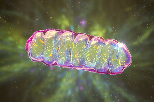
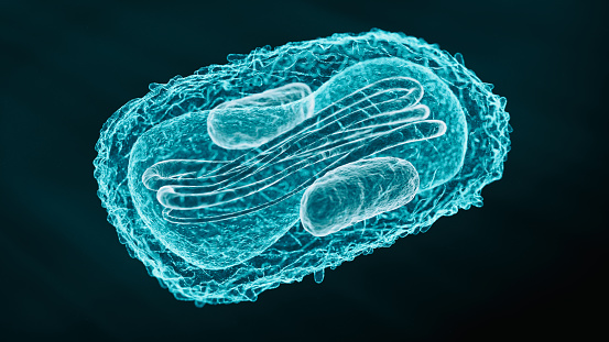
Free Images: "bestof:Animal mitochondrion diagram en (edit).svg A diagram showing a mitochondrion of the eukaryotic cell Mitochondria are organelles surrounded by membranes"
Diagram_of_the_Federal_Government_and_American_Union_edit.jpg
Animal mitochondrion diagram en (edit).svg
Animal mitochondrion diagram unlabelled.svg
Animal mitochondrion diagram en.svg
Animal mitochondrion diagram it.svg
Endomembrane system diagram en (edit).svg
Animal mitochondrion diagram bn.svg
Animal mitochondrion diagram es.svg
Animal mitochondrion diagram gl.svg
Animal mitochondrion diagram fi.svg
Animal mitochondrion diagram de.svg
Animal mitochondrion diagram hu.svg
Animal mitochondrion diagram pt.svg
Animal mitochondrion diagram fr.svg
Animal mitochondrion diagram pl.svg
Animal mitochondrion diagram lt.svg
Animal mitochondrion diagram zh hk.svg
Animal mitochondrion diagram zh tw.svg
Animal mitochondrion diagram ml.svg
Animal mitochondrion diagram zh ml.svg
Animal mitochondrion diagram cs.svg
Animal mitochondrion diagram ru.svg
Animal mitochondrion diagram uk.svg
Animal mitochondrion diagram id.svg
Animal mitochondrion diagram hi.svg
Animal mitochondrion diagram ca.svg
Eukaryotic cilium diagram en.svg
Mitochondrion mini.svg
Mitochondrion (numbers version).svg
Mitochondrion_(borderless_version)-pt.svg
Cellular tight junction-en.svg
Animal Cell.svg
Plant cell structure svg labels.svg
Cellular tight junction-fr.svg
Eukaryotic cilium diagram pl.svg
Diagram human cell nucleus cs.svg
Animal cell cycle-en.svg
Endomembrane system diagram en (edit) gl.svg
Complete neuron cell diagram tr.svg
Animal cell structure en.svg
Animal cell structure en gl.svg
Cell membrane detailed diagram pl.svg
Endomembrane system diagram en.svg
Gray5.svg
Animal cell structure gl.svg
Commercial_Hunter_Cell_Model.svg
Gray5_-_es.svg
Outer hair cell and Deiter's cell.svg
Bilayer hydration profile.svg
Desmosome cell junction pl.svg
Animal cell cycle (editors version).svg
Lipid unsaturation effect.svg
Gap cell junction keys.svg
Cellular tight junction-it.svg
Ribossomo tradução mRNA.svg
Gray5-ja.svg
Ribosome mRNA translation zh.svg
Cellular tight junction keys.svg
Ribosome mRNA translation bg.svg
Ribosome mRNA translation it.svg
Animal cell structure de.svg
Animal cell structure no text.svg
Animal cell structure pt.svg
Zelula prokarioto tipikoa.svg
Chloroplast diagram.svg
Apoplast and symplast pathways Dutch txt.svg
Animal cell cycle-zh-hant.svg
Mendel seven characters es.svg
Gray769-pt.svg
Animal cell structure sv.svg
Lipid unsaturation effect es.svg
Gray5_de.svg
Gray5_nl.svg
Chloroplast diagram (zh-cn).svg
Animal cell structure id.svg
Schema ciglio cellula eucaritica..svg
Ribosom mRNA translation de.svg
Mitochondrion (standalone version)-en.svg
Mitochondrion (borderless version)-en.svg
Mitochondrion structure labels.svg
Mitochondrion structure drawing.svg
Eukaryotic cilium diagram it.svg
Diagram human cell nucleus.svg
Prokaryote cell diagram he.svg
Diagram human cell nucleus sk.svg
Cell membrane detailed diagram en.svg
Prokaryote cell diagram cs.svg
Prokaryote cell diagram international.svg
Diagram human cell nucleus tr.svg
Plant cell wall diagram-en.svg
Complete neuron cell diagram en.svg
Mitochondrion (editors version).svg
Cell membrane detailed diagram et.svg
Digestive system diagram edit lv.svg
Diagram human cell nucleus sl.svg
Digestive system diagram edit lt.svg
Culex_pipiens_diagram_en.svg
Cell membrane detailed diagram es.svg
Complete neuron cell diagram he.svg
Cell membrane detailed diagram id.svg
Terms of Use   Search of the Day