Click Here for More Images from iStock- 15% off with coupon 15FREEIMAGES 
Blue colored stem cells of a lentil plant under the microscope.
plant Flower bud of Lilium brownii var.viridulum T.S.\nunder light mircoscope with white background
The study of plant tissues under the microscope in the laboratory.
corn stem micrograph with dye
Epidermis. Electron microscope micrograph showing a keratinocyte of spinous layer. The epithelial cell has a polygonal shape, central nucleus with nucleolus, cytoplasm full of keratin filament bundles, and numerous dark desmosomes crossing the intercellular spaces.
Photomicrograph of
centropyxis aculeata (amoeboid organism) under the microscope - optical microscope x400 magnification
Particles, medical concept
Components of Eukaryotic cell, nucleus and organelles and plasma membrane - 3d illustration
plant rape flower cross section under light mircoscope with white background
Leiden leaf c.s. under microscope
Pleomorphic liposarcoma. Site: Neck. Pleomorphic liposarcoma is a rare subtype of fat cell cancer in the body. It is a rare and very malignant type of tumor that usually arises from fat cells and deep soft tissues of the upper and lower extremities (shoulder, arms, legs, and thighs).
Cross-section Dicot, Monocot and Root of Plant Stem under the microscope for classroom education.
Colorful seamless pattern with Crystal stones.
FNAC of lymph nodes tissue Cytology microscopic 100x show Microbacterium Tuberculosis TB. Lymph node TB at medical histopathology laboratory.
Viral Infection Monkeypox Virus - 3d rendered image. Abstract biomedical illustration. \nAntibody, Antigen, Vaccine technology concept.
Microscope photo of a sambucus stem with parenchyma cells.
Digitally painted on Tablet, Pixelated Planets, Abstract Background
Freshwater Closterium algae (unicellular charophyte green algae) - optical microscope x200 magnification
Photomicrograph of two rotifers. Rapidly rotating cilia at top of heads, internal organs visible. Live specimen. Wet mount, 10X objective, transmitted brightfield illumination.
Enlarge microscopic view of larvae stage of crustacean
Microscopic image of dog stomach cardiac region
Eggs of helminth and Finding parasites in feces, analyze by microscope in laboratory.
Micro world
The algae species Pediastrum boryanum under the microscope.
Diffuse large B-cell lymphoma, or DLBCL, is a cancer that starts in white blood cells called lymphocytes.
Corn ster c.s. under microscope
Natural agate surfaces, suitable for backgrounds and wallpapers.
Cysts of Entamoeba coli protozoan, 3D illustration. E. coli is a non-pathogenic ameba, its cyst is 15-25 mkm, has 8 nuclei and chromatoidal bar elongated with splintered ends (shown red)
Intricate bubble texture macro in bright colors for abstract background. Liquid bubbles lit from below with colors injected into the liquid as a design or art abstract.
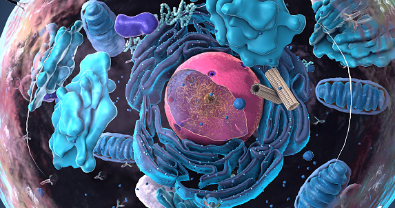
Free Images: "bestof:Animal cell structure ca.svg validSVG ca Diagrama d'una cel·lula animal 2015-04-30 File Animal_cell_structure_en svg User LadyofHats Translated by User"
Animal cell structure ca.svg
Animal cell structure id.svg
Animal cell structure no text.svg
Animal mitochondrion diagram cs.svg
Animal cell structure sv.svg
Animal mitochondrion diagram id.svg
Animal mitochondrion diagram ml.svg
Animal mitochondrion diagram zh ml.svg
Animal mitochondrion diagram zh hk.svg
Animal mitochondrion diagram zh tw.svg
Animal mitochondrion diagram en.svg
Animal mitochondrion diagram bn.svg
Animal mitochondrion diagram en (edit).svg
Acrosome reaction diagram es.svg
Prokaryote cell-ro.svg
Average prokaryote cell- nl.svg
Animal cell structure fr.svg
Anima cell notext.svg
Animal cell cycle-es.svg
Plant cell structure svg zh-hant.svg
16CygBbOrbit-ca.svg
Solar eclipse ca.svg
Plant cell structure svg-id.svg
Desmosome cell junction it.svg
Animal mitochondrion diagram it.svg
Animal mitochondrion diagram ru.svg
Endomembrane system diagram nl.svg
Endomembrane system diagram ln.svg
Main protein structure levels bg.svg
Main protein structure levels zh.svg
Average prokaryote cell cs.svg
Main protein structure levels pl.svg
Zelula prokarioto tipikoa.svg
Scheme simple diffusion in cell membrane-ca.svg
Scheme facilitated diffusion in cell membrane-ca.svg
Scheme sodium-potassium pump-ca.svg
Animal cell structure en.svg
Animal cell structure hu.svg
Animal cell structure pl.svg
Animal mitochondrion diagram ca.svg
Animal cell structure cs.svg
portugues-sexo_alemão-sex_ingles-sex_africano-seks_russo-секс_albanes-sex_chines-性別_bulgaro-секс_croata-spol_gales-rhyw_coreano-섹스_frances-sexe_japones-セックス_filandes-sukupuolesta_arabe-الجنس_prontos_ai_tens_em_15_linguas_a_p.jpg
Insulin glucose metabolism ZP-ca.svg
Animal cell structure gl.svg
Animal cell structure de.svg
Animal cell structure tr.svg
Animal Cell Unannotated.svg
Cell junction simplified zh traditional.svg
4 point lighting-ca.svg
Desmosome cell junction en.svg
Cell junction simplified it.svg
Animal cell cycle-en.svg
Turgor pressure on plant cells diagram.svg
Osmotic pressure on blood cells diagram.svg
Animal mitochondrion diagram unlabelled.svg
Function machine2-ca.svg
Cell junction simplified pt.svg
Erythrozyten und Osmotischer Druck.svg
Plant cell structure no text-2.svg
Cell membrane detailed diagram id.svg
Plant cell wall diagram mk.svg
Cell membrane detailed diagram ca.svg
Average prokaryote cell- id.svg
Mature flower diagram-ca.svg
Endomembrane system diagram ka.svg
Endomembrane system diagram zh.svg
Endomembrane system diagram en.svg
Osmotic pressure on blood cells diagram pl.svg
Osmotic pressure on blood cells diagram-es.svg
Plant cell structure svg 2.svg
Wikipedia article-creation-2-ca.svg
Scheme simple diffusion in cell membrane-zh (zh-tw).svg
Scheme sodium-potassium pump-el.svg
Canadian flag construction sheet-he.svg
Scheme sodium-potassium pump-he.svg
Turgor pressure on plant cells diagram (zh-tw).svg
Scheme secundary active transport-heb.svg
Canadian flag construction sheet-he2.svg
San Jose-San Francisco-Oakland CSA.svg
Flag of Canada and Austria.svg
Flag of Canada and Italy.svg
Flag of Canada and Germany.svg
Animal Cell.svg
Animal cell structure pt.svg
Animal cell structure uk.svg
Animal cell structure numbered version.svg
Animal cell structure en gl.svg
Iberian Peninsula in 125-en.svg
Complete neuron cell diagram ca.svg
Animal cell cycle-zh-hant.svg
Flag of French language.png
Scheme ant worker anatomy-ca.svg
Animal mitochondrion diagram fr.svg
Animal mitochondrion diagram hu.svg
Animal mitochondrion diagram pl.svg
Animal mitochondrion diagram hi.svg
Desmosome cell junction de.svg
Plant cell structure svg-zh-cn.svg
Terms of Use   Search of the Day