Click Here for More Images from iStock- 15% off with coupon 15FREEIMAGES 
Realistic 3D Render of Earthworm
plant Zea Stem C.S. under light mircoscope with white background
Epidermis. Electron microscope micrograph showing a keratinocyte of spinous layer. The epithelial cell has a polygonal shape, central nucleus with nucleolus, cytoplasm full of keratin filament bundles, and numerous dark desmosomes crossing the intercellular spaces.
Captured by a scientific microscope and Canon 5D Mark IV
Fingernail Cross Section of Tissue .
Photomicrograph of filamentous blue-green algae, Oscillatoria species. Each band along the stand is one cell. This filament is very short probably having recently parted from a longer one. Filaments are typically many times longer. This algae is mobile, it moves and twists through the water. San Francisco Bay, California, USA. Live specimen. Wet mount, 40X objective, transmitted brightfield illumination. Note - motion blur of live specimen, very shallow depth of field, chromatic aberration and uneven focus are inherent in light microscopy.
Backgrounds of Characteristics Tissue Tongue, Small intestine of Cat and Tissue of Esophagus rabbit under microscope.
Blastomycosis is a fungal infection caused by inhaling Blastomyces dermatitidis spores. If it involves only the lungs, it is called pulmonary blastomycosis. Only about half of people with the disease have symptoms, which can include fever, cough, night sweats, muscle pains, weight loss, chest pain, and feeling tired.
mosquito larvae (chironomus sp.) under the microscope - light microscope x32 magnification
Liver Fluke Section under light microscope with white background
Leiden leaf c.s. under microscope
Close-up view of the background honeycomb texture. Useful to help add freshness, sweet aroma from natural honeycomb, increase energy, and strengthen the body's immunity.
earth worm isolated on white background
Photomicrograph of planaria, a flatworm, Dugesia species. Slight motion blur in head. Freshwater stream, San Luis Obispo, California. Live specimen. Wet mount, 2.5X objective, transmitted brightfield illumination.
The detrusor muscle remains relaxed to allow the bladder to store urine, and contracts during urination to release urine 3d illustration
Diffuse large B-cell lymphoma, or DLBCL, is a cancer that starts in white blood cells called lymphocytes.
Internal structure of an animal cell, 3d rendering. Section view. Computer digital drawing.
plant rape flower cross section under light mircoscope with white background
Study of Parasitic helminths (Trematodes) and Ascaris of fishes under a microscope.
Isolated on white model of normal human skin with hair.
Hand Drawn of Human Anatomy Temporal Bones on Chalkboard
earth worm in soil
amazing inhabitants of the microworld under a microscope
Stinging nettle thorn under microscope
Asian swamp eel Monopterus albus isolated on white background
Pheochromocytoma, Site: Adrenal gland
Human hair under microscope, 3D illustration showing close-up structure of healthy human hair
Photomicrograph of segmented worm from salt marsh in San Francisco Bay, California, USA. Digestive system and other internal organs clearly visible. Live specimen. Wet mount, 2.5X objective, transmitted brightfield illumination.
Plant tissue Structure, section (tissue) of stem plant tissue under a light microscope.
3d Mitochondrion isolated on black
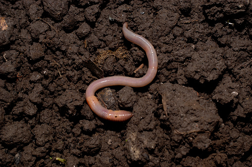
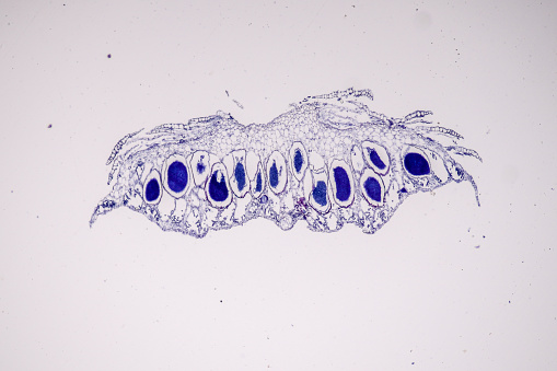
Free Images: "bestof:Anatomy of an earthworm 001.png en Cross section of the body of an earthworm Oligochaeta showing the disposition of the more important organs the body wall w"
mummification-skeleton-horse-reiter-60510.jpg
Secondary tumor deposits in the liver from a primary cancer of the pancreas.jpg
anatomy-woman-human-face-body-254120.jpg
anatomy-man-human-face-body-lips-254129.jpg
fossil-petrification-fish-skeleton-201044.jpg
woman-human-pair-man-face-body-254133.jpg
road-construction-road-romans-roman-246931.jpg
road-construction-road-romans-roman-246929.jpg
Human anatomy.jpg
Naked human male body front anterior.png
Naked human male body front anterior (annotated-id).png
Persons.jpg
John-bell-II-B-6.jpg
Human anatomy ru 2.jpg
Haeckel_Calcispongiae.jpg
Anatomía humana.png
8._desember_-_December_8.jpg
P6_Utanka_and_The_Earrings.png
Cross section jellyfish en.svg
Jellyfish cross section numbered.svg
Parborlasia_corrugata_body_wall.svg
Gray1167.svg
Стомах.svg
Gray1193.svg
Gray1046.svg
Gray1167_fi.svg
Gray5.svg
Gray1167 es.svg
Gray5-ja.svg
Gray1046_pl.svg
Gray1236_pl.svg
Gray799.svg
Gray1193_pl.svg
Gray1235_pl.svg
Gray5_-_es.svg
Gray769-es.svg
Gray769-pt.svg
Gray1167_pl.svg
Gray5_de.svg
Gray5_nl.svg
Cross section jellyfish ru.svg
Gray67.svg
Design_Analysis_of_the_P-38_Lightning.png
Schwarzschild cross section.svg
Internal organs.svg
Bilaterian-plan.svg
Bilaterian-plan hy.svg
Gray1130.svg
Gray728.svg
Gray727.svg
Gray477.svg
Gray399.svg
Gray38.svg
Gray726.svg
Gray1199_pl.svg
Gray37.svg
Gray834.svg
Gray726-Brodman.svg
Gray726-Brodman-prefrontal.svg
Gray399_es.svg
Gray63.svg
Gray1199_en.svg
Gray511.svg
Gray506_es.svg
Gray770-es.svg
Gray770-pt.svg
En-elbow joint.svg
Gray1167_de.svg
Gray1216.svg
Gray506_pl.svg
Gray1095_-_pl.svg
Temporo-parietal_junction_ja.svg
Gray506-ar-p.svg
Culex_pipiens_diagram_en.svg
Gray727_el.svg
Gray1199_labeled.svg
Gray511-es.svg
Gray834_es.svg
Ant worker morphology (ja).svg
Gray812and814_es.svg
Lobes_of_the_brain_el.svg
Gray512.svg
Gray1216_modern_locations.svg
Trilobite sections numbered.svg
Culex pipiens diagram es.svg
Gray814_pl.svg
Gray1144.png
Gray1095.svg
12._desember_-_December_12.jpg
George_Stubbs_-_A_Comparative_Anatomical_Exposition_of_the_Structure_of_the_Human_Body_with_that_of_a_Tiger_and_a_Co..._-_Google_Art_Project_(2348692).jpg
George_Stubbs_-_A_Comparative_Anatomical_Exposition_of_the_Structure_of_the_Human_Body_with_that_of_a_Tiger_and_a_Co..._-_Google_Art_Project_(2349640).jpg
Scheme ant worker anatomy-en.svg
Dragonfly.svg
George_Stubbs_-_A_Comparative_Anatomical_Exposition_of_the_Structure_of_the_Human_Body_with_that_of_a_Tiger_and_a_Co..._-_Google_Art_Project_(2347745).jpg
lungs-diagram-anatomy-body-human-41562.svg
man-woman-schema-body-anatomy-144378.jpg
Scheme_body_cavities-en.svg
Aura body.svg
Oligochaeta anatomy.svg
Human body concentrations.svg
Terms of Use   Search of the Day