Click Here for More Images from iStock- 15% off with coupon 15FREEIMAGES 
MRI of spinal column
An MRI of the whole spine image is a comprehensive visual representation produced through Magnetic Resonance Imaging, providing detailed insights into the entire spinal structure.
Businessman with backache against white background.
The lumbar vertebrae are, in human anatomy, the five vertebrae between the rib cage and the pelvis .
Doctor with a pen showing a hernia in a skeleton to a patient concept
Cervical Vertebrae Bridge X-ray Film
cervical spine xray from xray results of pelvic injury. Real cervical spine xray from hospital. cervical spine xray due to impact
Spinal Cord X-ray Film
Male doctor holding and viewing x-ray.
CT SCAN of Chest and Abdomen Coronal and sagittal view with injection contrast media for diagnosis chest and abdomen disease.
Protecting patient`s health and recovery concept. Neutral color palette, copy space for text.
Scoliosis spine x-ray of a woman body
The lumbar vertebrae are, in human anatomy, the five vertebrae between the rib cage and the pelvis.
waist pain three Dimensional
Diagnosis of spinal diseases using computed tomography.
Female doctor talking about spinal cord problems
African descent doctor looking x-ray image on digital tablet. Isolated on white background. Shoot with Canon EOS 5DS full frame DSLR.
The nuchal ligament is a ligament at the back of the neck that is continuous with the supraspinous ligament 3d illustration
Real Tomography Diagnostic of patient with osteoporosis, pain in the spine. Magnetic Resonance Imaging MRI scanner Radiology. Computed x-Ray Medical Imaging
Computational tomography image of the abdominal and pelvic regions.
The vertebral column is composed of 33 vertebrae, divided into five regions: cervical (7), thoracic (12), lumbar (5), sacral (5 fused into the sacrum), and coccygeal (4 fused into the coccyx). It provides support and protects the spinal cord.
Xray of ones manhood.
C-spine x-ray or Cervical spine image lateral views of female patient for doctor diagnosis on intervertebral disc herniation or Cervical radiculopathy, Spondylosis and fracture
Human neck X-ray Film
Male doctor examining X-Ray image isolated over white background
In vertebrates, thoracic vertebrae compose the middle segment of the vertebral column, between the cervical vertebrae and the lumbar vertebrae 3d illustration
Backbone and joint diagnostics. Lumbar and skeleton test.
Xray film of a patient with scoliosis of lumbosacral spines
Collection of MRI of whole spine  T2W sagittal  plane for diagnostic Spinal Cord Compression.
Cogs marks on a spray-painted surface.
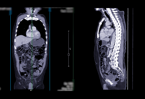
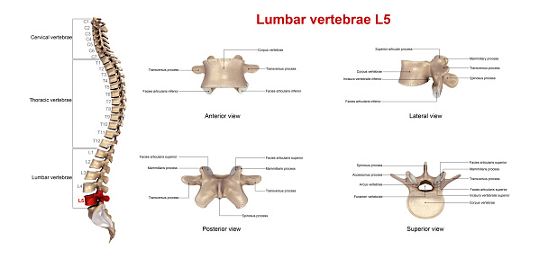
Free Images: "bestof:ARACHNOIDITIS.JPG en Myelogram showing arachnoiditis in the lumbar spine Own A E Francis 1/16/09 Myelography"
ARACHNOIDITIS.JPG
CT ARACHNOIDITIS.JPG
Spinal stenosis 1.JPG
SPINAL_STENOSIS.JPG
CT LUMBAR DISC HERNATION.JPG
Stmarygamul.jpg
LAMINECTOMY SCAR.JPG
WestinStFrancis.jpg
L1 2 vertebral fracture.jpg
Anne de Bretagne as prudence.jpg
Antes01.png
Antes02.png
Beaver wars map.jpg
White serbia white croatia01.png
White serbia white croatia02.png
StromerzgFrancisTurbine.jpg
Spinal-disc-protrusion-l5.jpg
RichardIdeGrenville_KilkhamptonChurch_1860.JPG
RobertFitzHamon_&RichardDeGrenville_1860Window_Kilkhampton.JPG
Terms of Use   Search of the Day