Click Here for More Images from iStock- 15% off with coupon 15FREEIMAGES 
Nirmatrelvir inhibits the 3C-like protease of the SARS-CoV 2 virus. The coronavirus 3CL hydrolase enzyme, also known as the main protease, is essential for the maturation of the virus.
Mitochondrial cardiomyopathy. Site: Heart. Mitochondrial diseases are a clinically and genetically heterogeneous group of disorders that result from dysfunction of the mitochondrial respiratory chain, which is responsible for the generation of most cellular energy.
3D cartoon and Gaussian surface models, PDB 1uea, white background
A molecular model of Human Insulin. Isolated on black.
Cytochrome P450 molecule. CYP 3A4 is the most promiscuous of the human CYP enzymes. Molecular model. 3D rendering. Illustration
Inside protein molecule
Cediranib cancer drug molecule. 3D rendering. Atoms are represented as spheres with conventional color coding.
Freshness capsule
Bacterial multidrug efflux pumps (E-CmeB) expel antibiotics  from inside the bacterium (bottom): ciprofloxacin (yellow), chloramphenicol (violet),  ampicillin (red).
Wilms tumor (also called Wilms' tumor or nephroblastoma) is a type of childhood cancer that starts in the kidneys. It is the most common type of kidney cancer in children.
3D cartoon and Gaussian surface models, PDB 7bg9, white background.
Liver Fluke Section under light microscope with white background
Collagenase clostridium histolyticum, an enzyme produced by the bacterium Clostridium histolyticum that dismantles collagen. Used as an injection kit for the treatment of Dupuytren's contracture.
Chemical formula of Progesterone
Acetylcholine receptors in the lipid layer of cell membrane.
The three-dimensional structure of proteins is represented in a variety of conformations. This image is based on the ribbon structure of a protein. It is suitable for biology notebooks and book covers.\nThis image has not been created by AI.
PETase is a bacterial enzyme that breaks down PET-plastic to monomeric molecules. The whole bacterial degradation process yields terephtalic acid and ethylene glycol, which are environmentally harmless.
Diffuse large B-cell lymphoma, or DLBCL, is a cancer that starts in white blood cells called lymphocytes.
3D cartoon and Gaussian surface models, chain id color scheme, PDB 2v0z, white background
plant Stem of Nymphaea tetragona T.S. under light mircoscope with white background
Green algae cells background
Photomicrograph of filamentous blue-green algae, Oscillatoria species. Each band along the stand is one cell. This filament is very short probably having recently parted from a longer one. Filaments are typically many times longer. This algae is mobile, it moves and twists through the water. San Francisco Bay, California, USA. Live specimen. Wet mount, 40X objective, transmitted brightfield illumination. Note - motion blur of live specimen, very shallow depth of field, chromatic aberration and uneven focus are inherent in light microscopy.
Drops and strands of hot glue have already cooled down and are visible against a dark blue background
Liquid bubbles up Fluid Twirl
PETase (left) degrades  polyethylene terephtalate (PET) into monohydroxyethyl terephthalate (MHET) and MHETase degrades MHET ethylene glycol (EG) and terephthalic acid (TPA).
Fatty liver disease (steatosis) is a common condition caused by having too much fat build up in your liver. A healthy liver contains a small amount of fat. It becomes a problem when fat reaches 5% to 10% of your liver's weight.
3D cartoon and Gaussian surface models, PDB 6gu2, white background
Dessert
Coxsackie virus internal structure, 3d illustration set isolated on white
Molecular Model of Vitamin E.
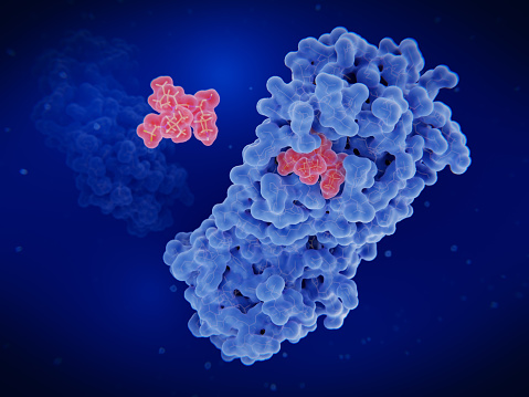
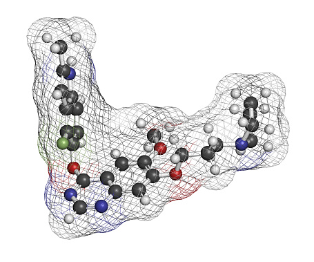
Free Images: "bestof:AA in COX-2.png en Archidonic Acid bound to the COX-2 enzyme own Ppruks 2011-05-16 Cc-zero Enzymes"
Cox-2010-stl.jpg
Papoose sunset.jpg
Monarch Sunrise Wing.jpg
Monument 151 de Tonina, prisonnier, exposition "Mayas", Musée du Quai Branly, Paris.jpg
Retablo virgen mareantes 1 Alcazar Seville Spain.jpg
BD Cox's Bazar District locator map.svg
Etoricoxib structural formula V.1.svg
Janet Cox-Rearick Waldman with portrait of Eleonora di Toledo by Agnolo Bronzino.jpg
P450cycle.svg
Mecanism of action for Lysozyme.svg
Tryptanthrin.svg
Rofecoxib.svg
TitanicAcidNanotubeStructure.svg
2-(aminooxy)aceticacid2.svg
Induced fit model en.svg
Fluoroacetic Acid V.1.svg
Τιτλοδότηση.svg
Rhodanase2 V.1.svg
Eudesminsäure.svg
Sulfanilic+H2SO4+NaNO2.svg
Chlorogenic-acid.svg
Phosphoenolpyruvic acid Structural Formulae V.1.svg
Folic acid (2).svg
Ornithursaeure.svg
Isobutyric acid Formula V.1.svg
Pangamic acid Structure V.1.svg
Acromelic acid A structure.svg
Cerebronic acid.svg
Citronellic acid.svg
Ultimate pictogram.svg
Gadoleinsäure.svg
Tribromoisocyanuric acid.svg
Ginkgolic acid 2.svg
Polyhydroxyacetic Acid V.1.svg
Linoleic Acid Structural Formulae V.1.svg
Linoleic Acid Structural Formulae V.2.svg
Chelidonic acid Structural Formula V.1.svg
Diphenic Acid Structural Formula V.1.svg
Acetic Acid Hydrogenbridge V.2.svg
Cucurbinic Acid Structural Formula V.1.svg
Acromelic acid B structure.svg
2-Fluoroacrylic acid.svg
Portrait of Henry L. Fry by Kenyon Cox, Cincinnati Art Museum.JPG
Alpha-Linolenic acid Structural Formulae V.2.svg
Punicic Acid Structural Formulae V.1.svg
Alpha-Linolenic acid Structural Formulae V.1.svg
Methyl acrylate V.1.svg
Cyano Acetic Acid Formula V.1.svg
2,4,5-trimethoxybenzoic acid.svg
Acetic Acid Hydrogenbridge V.1.svg
Bromofluorosulfate structure.svg
Citric acid cycle ru.svg
Louise_Howland_King_Cox,_A_Rondel,_1892,_oil_on_canvas,_High_Museum_of_Art,_Atlanta,_Georgia.jpg
Oelic Acid Structural Formulae V.2.svg
Mycophenolic acid Strucrural Formulae V.1.svg
Undeca-2E-en-8,10-diynoic acid isobutylamide.svg
Cerotic acid.svg
Peracetic Acid Structural Formula V.1.svg
(R)-Mosher Acid Formula V.1.svg
(S)-Mosher Acid Formula V.1.svg
Diphenic Acid Synthesis V.1.svg
Nalidixic acid structure.svg
Formic Acid Hydrogenbridge V.1.svg
Flufenamic acid synthesis.svg
Synthesis of carbamoyl aspartic acid.svg
Pivalic Acid Structural Formulae V.1.svg
Ether-cleavage-sn1.svg
Ether-cleavage-sn2.svg
Amino Sulfonic Acid Synthesis V.1.svg
Sodiumsalt of thioglycolic acid V.1.svg
Ammoniumsalt of thioglycolic acid V.1.svg
Nalidixic acid synthesis 01.svg
Prosztánsav.svg
Chlorofluorosulfate structure.svg
Mefenamic acid synthesis 01.svg
Secalonic acid D or ergochrome EE.svg
Diana by Kenyon Cox, c. 1890, oil on canvas - Chazen Museum of Art - DSC02161.JPG
Glutamate dehydrogenase reaction.svg
Degradation of uric acid.svg
Na2CO3 HCl titration.svg
Oelic Acid Structural Formulae V.1.svg
Folic acid nglu.svg
(R)- & (S)-Mosher Acid Formula V.1.svg
Mesoxalic acid hydration equilibrium.svg
Orthanilic acid.svg
Myristoleic acid-1.svg
Types of inhibition sr.svg
Secalonic acid B or ergochrome BB.svg
Secalonic acid C or ergochrome AB.svg
Approach of Love by Kenyon Cox, 1890-1893, oil on canvas - Cincinnati Art Museum - DSC04496.JPG
Magnesium valproate.svg
Mosher's Reagent Formulae V.1.svg
8-cube t34.svg
Titration weakbase strongacid.svg
Iopanoic Acid Structural Formulae V.1.svg
Vapour pressure trifluoroacetic acid.svg
Propionic acid chemical structure.svg
Sodium butyrate formula V.1.svg
O-Ethyl methylphosphonothioic acid Structural Formula V.1.svg
Aspartate transaminase reaction.svg
Terms of Use   Search of the Day