Click Here for More Images from iStock- 15% off with coupon 15FREEIMAGES 
From left to right: the NMDA and  AMPA receptors transport calcium cations into neurons after being activated by the neurotransmitter glutamate and  the GABA receptor (right) transport chloride anions after the activation by gamma aminobutyrate. PDB entries: 6wht,  3kg2, 6d6u
The three-dimensional structure of proteins is represented in a variety of conformations. This image is based on the ribbon structure of a protein. It is suitable for biology notebooks and book covers.\nThis image has not been created by AI.
3D cartoon and Gaussian surface models, chain id color scheme, PDB 7t3a, white background
A ball and stick model of Human Insulin. Isolated on black.
Proteasomes are molecular machines for breaking down proteins called proteolysis, only target proteins that have been marked for destruction called ubiquitin to the target protein, 3d rendering
Inside protein molecule
Cell Membrane Phospholipid Structure,Phospholipid bilayer of the cell membrane,Lipid bilayer
Multicolored antibodies or immunoglobulin protein structures - 3d illustration
Proteasomes are large molecular machines that degrade unneeded or damaged proteins that have been tagged with polyubiquitin.
Endosomes - 3d rendered image of Endosomes are membrane-bound organelles. They are parts of endocytic membrane transport pathway.  Endomembrane system in cells. Bilayer Lipid Membrane. Abstract medical illustration. Microscopic view. Microbiologic research concept.
Cell membrane and biology, biological concept, 3d rendering. Computer digital drawing.
Covid-19 Coronavirus Variants Omicron Delta Gamma Beta Alpha Pandemic Virus Disease Mutation
DNA close-up, DNA Molecule Model, DNA Strands, DNA animation
3D Rendered Illustration, visualization of Neurons firing neurotransmitters in the synaptic gap.
3D cartoon and Gaussian surface models, chain id color scheme, PDB 3omv, white background
Cells in the body examined by endoscope
GABA B receptors  are G protein-coupled receptors, also called metabotropic receptors.  Binding of  an agonist (baclofen, red) leads to a G-protein coupled C-AMP signaling pathway. Source: PDB entries 7eb2, 6r3q,.
Endosomes - 3d rendered image of Endosomes are membrane-bound organelles. They are parts of endocytic membrane transport pathway.  Endomembrane system in cells. Bilayer Lipid Membrane. Abstract medical illustration. Microscopic view. Microbiologic research concept.
Collagen protein molecule. Molecular model. 3D rendering. Illustration
A ball and stick + ribbons model of beta-secretase - a protein that is important in forming healthy peripheral nerve cells, but is also involved in the onset of Alzheimer's disease.
c-Myc and Max transcription factors bound to DNA. 3D illustration. Atoms shown as spheres with conventional color coding.
DNA A-T Pair
3D cartoon and Gaussian surface models, PDB 6rwb, white background
nanotechnology and abstract graphene structures
The glucagon like peptide-1 receptor (GLPr) is a G proteincoupled receptor (GPCR) that mediates the action of GLP-1, a peptide hormone. The activated receptor  has a strong effect  on the management of type 2 diabetes mellitus and obesity, including glucose homeostasis and regulation of gastric motility and food intake.
Endosomes - 3d rendered image of Endosomes are membrane-bound organelles. They are parts of endocytic membrane transport pathway.  Endomembrane system in cells. Bilayer Lipid Membrane. Abstract medical illustration. Microscopic view. Microbiologic research concept.
organic chemistry, model of the protein molecule - illustration of a biological particle
Focal nodular hyperplasia (FNH) is a benign liver lesion that is composed of a proliferation of hyperplastic hepatocytes surrounding a central stellate scar. Typically, FNH is a solitary lesion that is more commonly seen in women.
3d rendered Gastric inhibitory polypeptide (GIP, glucose-dependent insulinotropic peptide) endocrine protein hormone, 3D illustration
Artificial Intelligence Sustainable Responsible Technology
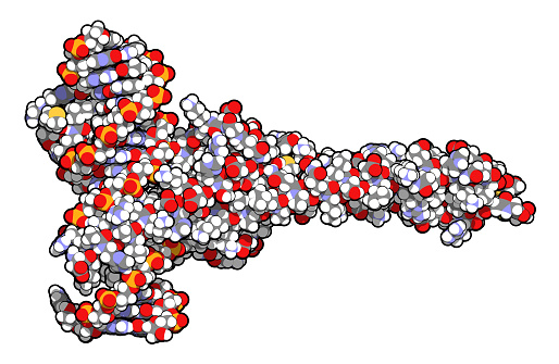
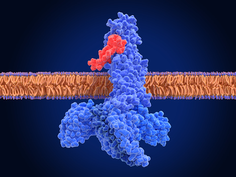
Free Images: "bestof:A2A receptor bilayer.png 3EML coordinates Own Boghog2 2008-10-15 Protein structures G protein coupled receptors"
Protein 7 domen.svg
ZM241385.svg
Conserved residues.svg
Sumatriptan binding site.svg
ORs and GP.svg
Main protein structure levels en.svg
Main protein structure levels pl.svg
Main protein structure levels fr.svg
Main protein structure levels gl.svg
Main protein structure levels zh.svg
Main protein structure levels bg.svg
Steatosis.svg
Aa structure function.svg
Molbio-Header-2.svg
Molbio-Header.svg
A2A receptor bilayer.png
NAchR 2BG9.png
PPARg RXRa 3E00.png
5ht2a.jpg
MDR3 3g5u.png
Reverse Transcriptase 1HMV.png
Thymosin 1HJ0.png
HTR2A Human 5-hydroxytryptamine receptor 2A.png
2LNL.pdb.png
3RZE.png
4DAJ.png
3V2Y.png
3VW7.png
4GRV.png
GPCR mechanism.png
7TM4 (GPCR).png
Beta-2 adrenergic receptor.png
PR DBD 2C7A.png
CNR1 Human Cannabinoid receptor 1.png
CNR1 HUMAN Cannabinoid receptor 1.jpg
Trpv1 pip2 bilayer.png
Trpv1 pip2 bilayer cropped.png
RORC 3L0L.png
Muscarinic acetylcholine receptor M2-3UON.png
GPCR activation.png
G-protein-coupled receptor.png
1ZHT.png
Prosaposin schematic.png
SSR149415.png
Rhodopsin 3D.jpeg
3RZE-human histamine H1 receptor-pdb.jpg
1BT6 tetramer.png
Bindisætiogsumatriptan12.PNG
Amibegron.png
Sorting nexin schematic.png
TMD.png
Transdução sinal adrenérgicos.PNG
Protein CNR2 PDB 2KI9.png
3ODU.png
ECL2 2RH1.png
4-NEMD.png
The effect of Rs and Gs in cAMP signal pathway.sr.png
The effect of Ri and Gi in cAMP signal pathway.sr.png
LDLR pathway.png
LDLR 1N7D.png
Modern protein hypothesis of mechanism of general anesthesia.png
PDGFR-beta 3MJG.png
Hexoprenaline.png
Beta2Receptor-with-Gs.png
ShK 1ROO.png
H1 Receptor with Doxepin.png
Steroid receptor.png
Type ii nuclear receptor action.png
E coli beta clamp 1MMI.png
Nodal signaling.jpg
1QBH BIRC2.png
2RH1.png
Determinants of Gastric Acid Secretion.png
GABAA-receptor-protein-example.png
ZM241385.png
Gprotein-coupled receptor kinase.png
PDB 1suv EBI.jpg
4DJH.png
PDB 1ld4 EBI.jpg
PDB 1m4x EBI.jpg
PDB 1w8x EBI.jpg
PDB 1qgc EBI.jpg
PDB 1dyl EBI.jpg
PDB 1hb7 EBI.jpg
PDB 1gw7 EBI.jpg
PDB 1hb5 EBI.jpg
PDB 1gw8 EBI.jpg
PDB 1hb9 EBI.jpg
PDB 1tzn EBI.jpg
PDB 1bm1 EBI.jpg
PDB 2brd EBI.jpg
PDB 1brd EBI.jpg
PDB 1m0k EBI.jpg
PDB 1m0l EBI.jpg
PDB 1m0m EBI.jpg
PDB 2ntu EBI.jpg
PDB 2ntw EBI.jpg
PDB 1p8h EBI.jpg
PDB 1kgb EBI.jpg
PDB 1f50 EBI.jpg
Terms of Use   Search of the Day