Click Here for More Images from iStock- 15% off with coupon 15FREEIMAGES 
A ball and stick model of Human Insulin. Isolated on black.
Bacterial multidrug efflux pumps (E-CmeB) expel antibiotics  from inside the bacterium (bottom): ciprofloxacin (yellow), chloramphenicol (violet),  ampicillin (red).
Proteasomes are molecular machines for breaking down proteins called proteolysis, only target proteins that have been marked for destruction called ubiquitin to the target protein, 3d rendering
The three-dimensional structure of proteins is represented in a variety of conformations. This image is based on the ribbon structure of a protein. It is suitable for biology notebooks and book covers.\nThis image has not been created by AI.
organic chemistry, model of the protein molecule - illustration of a biological particle
3d Rendering Molecular Structure,Ozone,Hydrogen and oxygen
A chromatosome is a result of histone H1 binding to a nucleosome, which contains a histone octamer and DNA.
background
Biological protein and molecule, 3d rendering. 3D illustration.
ATP Synthase Protein - 3d rendered image. Medical microbiology concept.  Structure and conformational states of the bovine mitochondrial ATP (Adenosine triphosphate) synthase. Chemical energy is synthesized in eukaryotic cells primarily. Hydrolase. Hologram view. HUD, FUI
A ball and stick + ribbons model of human Interleukin 2 or IL-2. Interleukin is a hormone which acts as a signalling molecule for the immune system.
Monosodium glutamate, MSG or sodium glutamate molecules. MSG is used in cooking as a flavor enhancer with an umami taste 3d rendering
p53 (blue) tagged with ubiquitin (yellow) is degraded into small peptides by a proteasome (violett). p53 prevents cancer formation and acts as a guardian of the genome. Mutations in the p53 gene contribute to about half of the cases of human cancer.
Artificial Intelligence Sustainable Responsible Technology
Deacetoxycephalosporin-C synthase (DAOCS) is a mononuclear ferrous enzyme that transforms penicillins into cephalosporins. 3D cartoon and Gaussian surface models, chain instance color scheme, based on PDB 1uof, white background.
Phospholipid bilayer membrane structures that make up living cells and their contents. This is an image of membrane proteins in the membrane.\nThis image, which looks like a plastic model, evokes biology and biochemistry. It can be used as a cover for magazines, textbooks and presentation materials.\nThis image was not created by AI.
Lab Experiment
Biological protein and molecule, 3d rendering. 3D illustration.
alien structure made of white spheres and flowing glass liquid, molecular, hi-tech concept, abstract design element
molecular structure digital concept
3d rendering of benzene molecules conjugated gold (Au) nanoparticles (NPs) on the black background
Inside protein molecule
3D cartoon and Gaussian surface models, chain id color scheme, PDB 7t3a, white background
Wireframe double helix DNA
Background of scientific cosmetic technologies. conceptual cosmetic solution for skin care. Background for biochemical, pharmaceutical, cosmetic and other medical concepts. Abstract science background. Abstract liquid, water. Molecules. 3D rendering.
H2 Hydrogen Molecule Fuel Cell Element Vehicle Biodegradable Ethical Renewable Energy
From left to right: the NMDA and  AMPA receptors transport calcium cations into neurons after being activated by the neurotransmitter glutamate and  the GABA receptor (right) transport chloride anions after the activation by gamma aminobutyrate. PDB entries: 6wht,  3kg2, 6d6u
3d rendering of Easter eggs background
Calcidiol molecule, also called calcifediol, major circulating metabolite of vitamin D3 produced in liver, 3D illustration
3D rendering of microscopic molecular structure
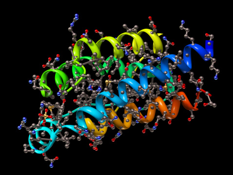
Free Images: "bestof:1ZHT.png Crystallographic structure of the oxysterol-binding protein based on the 1ZHT coordinates Own 2008-11-18 Boghog2 Ribbon diagrams"
Main protein structure levels en.svg
Main protein structure levels fr.svg
Main protein structure levels pl.svg
Aa structure function.svg
Milne universe with CMBR.svg
Animal cell structure en.svg
Animal cell structure de.svg
Euglenid pellicula scheme.svg
Main protein structure levels zh.svg
Main protein structure levels bg.svg
Monroe County Route 138 OH.svg
Monroe County Route 88 OH.svg
Articulacion malla.svg
Examples of Airfoils.svg
Complement pathway.svg
1ZHT.png
1BT6 tetramer.png
3OG7.png
Lactoperoxidase 2R5L.png
1CF1.png
CobB NAD dependent deacetylase 1S5P.png
IL22 IL22R 3DGC.png
1TIE.png
1QBH BIRC2.png
BLVRA 2H63.png
1A0A.png
3F5C.png
Reverse Transcriptase 1HMV.png
Acethylcholinesterase TC 1EA5.png
1R02 crystallography.png
1CQ0 crystallography.png
1SZD.png
FixL 1y28.png
EP300 3BIY.png
2LEF.png
3CNA Concanavalin A.png
1E5W.png
HSOD1 2VR6.png
1IC8.png
MDR3 3g5u.png
Thymosin 1HJ0.png
1BMC.png
1Y2A.png
1TU3.pdb.png
1HS6.png
HSP60 1SRV.png
31BI.png
1PWU.png
3RZE.png
Aromatase 3EQM.png
Kinesin motor domain 1BG2.png
1a3p egf.png
2e26.png
1H6F.png
3DYD.png
1ALK.png
2e52.png
1QPS.png
1RFX.png
1PEU R1E.png
NAchR 2BG9.png
4IC4.png
2ILA.png
ACD-TPP1 rasmol.png
Carbohydrate kinase 1KYH.png
BUB1 3E7E.png
EcKARI p-domain NADPH.png
E coli beta clamp 1MMI.png
PPARg RXRa 3E00.png
RORC 3L0L.png
1FG9 IFNG IFNG.png
2B8T.png
1T8U.jpg
LDLR Binding Module5 1ajj.PNG
Hsp60 structure.jpg
FhaC protein.GIF
2PX6 orlistat.png
Beta-2 adrenergic receptor.png
I-CreI dimer DNA 4.png
I-CreI dimer 1.png
I-CreI dimer DNA.png
I-CreI dimer DNA 2.png
2FO0 (c-Abl inactive).png
PDB 1zht EBI.jpg
A2A receptor bilayer.png
IL17F 1JPY.png
PGRP.png
1N6J.png
1efv opm.png
GHCS protein structure.jpg
1UVQ.png
SAP.png
Carnosine-2D-skeletal.png
1b9x opm.png
Structure cobl.jpg
POT1+adn rasmol.JPG
Cobatoxin 1 ribbon.png
Eif5 n.png
Eif5 c.png
Shh processing.png
Terms of Use   Search of the Day