Click Here for More Images from iStock- 15% off with coupon 15FREEIMAGES 
Colorful chain of amino acids or bio molecules called proteins - 3d illustration
The three-dimensional structure of proteins is represented in a variety of conformations. This image is based on the ribbon structure of a protein. It is suitable for biology notebooks and book covers.\nThis image has not been created by AI.
Proteasomes are molecular machines for breaking down proteins called proteolysis, only target proteins that have been marked for destruction called ubiquitin to the target protein, 3d rendering
A ball and stick model of Human Insulin. Isolated on black.
Biological protein and molecule, 3d rendering. 3D illustration.
Lab Experiment
Bacterial multidrug efflux pumps (E-CmeB) expel antibiotics  from inside the bacterium (bottom): ciprofloxacin (yellow), chloramphenicol (violet),  ampicillin (red).
Wireframe double helix DNA
3d helix chromosome or Dna structure, technology science background. 3d render illustration
dna helix
DNA close-up, DNA Molecule Model, DNA Strands, DNA animation
3d Rendering Molecular Structure,Ozone,Hydrogen and oxygen
A ball and stick + ribbons model of human Interleukin 2 or IL-2. Interleukin is a hormone which acts as a signalling molecule for the immune system.
3D cartoon and Gaussian surface models, chain id color scheme, PDB 7t3a, white background
Inhibition of mTORC1 by rapamycin leads to immunosuppression, prevention of trasplant rejection and inhibition of tumor growth.
Phospholipid bilayer membrane structures that make up living cells and their contents. This is an image of membrane proteins in the membrane.\nThis image, which looks like a plastic model, evokes biology and biochemistry. It can be used as a cover for magazines, textbooks and presentation materials.\nThis image was not created by AI.
Chain of amino acid or bio molecules called protein - 3d illustration
CRISPR-Cas9 gene editing technology
3d rendered Gastric inhibitory polypeptide (GIP, glucose-dependent insulinotropic peptide) endocrine protein hormone, 3D illustration
Inside protein molecule
Science background with molecule or atom
A ribbons model of an antibody, also known as immunoglobin. The immune system produces antibodies (using white blood cells) which then identify and attack viruses and bacteria.
3D cartoon and Gaussian surface models, PDB 6vg8, white background
background
Molecule with biology and chemical concept, 3d rendering. Digital drawing.
Molecular structure formula. Concept. 3D Render
p53 (blue) tagged with ubiquitin (yellow) is degraded into small peptides by a proteasome (violett). p53 prevents cancer formation and acts as a guardian of the genome. Mutations in the p53 gene contribute to about half of the cases of human cancer.
Hexagon honeycomb pattern metallic background, 3d render.
3D model of a protein molecule. Endothelial tyrosine kinase receptor, a marker of angiogenesis.
dna helix
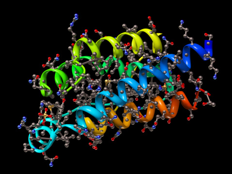
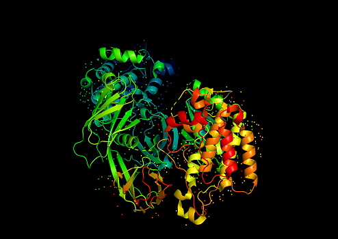
Free Images: "bestof:1PEU R1E.png Crystallographic structure of the ribonucleotide reductase protein R1E from Salmonella typhimurium The protein is rainbow colored N-terminus blue"
dna-biology-science-helix-protein-163710.jpg
1PEU R1E.png
RORC 3L0L.png
2OHV.png
EP300 3BIY.png
3EL3.png
1QBH BIRC2.png
FixL 1y28.png
1a3p egf.png
BLVRA 2H63.png
1HS6.png
1SZD.png
Aromatase 3EQM.png
Kinesin motor domain 1BG2.png
Rhodopsin-transducin.png
2PX6 orlistat.png
3CQV.png
PPARg RXRa 3E00.png
Crystal Structure of the Ku80 - 11.png
Crystal Structure of the Ku80 - 37.png
Crystal Structure of the Ku80 - 38.png
Crystal Structure of the Ku80 - 39.png
Crystal Structure of the Ku80 - 40.png
Crystal Structure of the Ku70 - 42.png
Crystal Structure of the Ku70 - 43.png
Crystal Structure of the Ku70 - 41.png
Crystal Structure of the Ku Heterodimer - 15.png
Crystal Structure of the Ku Heterodimer - 16.png
Crystal Structure of the Ku Heterodimer - 22.png
Crystal Structure of the Ku Heterodimer - 23.png
Crystal Structure of the Ku Heterodimer - 26.png
Crystal Structure of the Ku Heterodimer - 30.png
Crystal Structure of the Ku Heterodimer - 31.png
Crystal Structure of the Ku Heterodimer - 36.png
Crystal Structure of the Ku Heterodimer - 35.png
Mercuric reductase protein structure.jpg
1H6F.png
2e26.png
2HNX.png
G-Protein.png
1ZHT.png
1BT6 tetramer.png
3F5C.png
PBB Protein AKR1A1 image.jpg
2LEF.png
IL17F 1JPY.png
2RAM.png
PDB 1afr EBI.jpg
PDB 1oq9 EBI.jpg
PDB 1oq7 EBI.jpg
PDB 1otk EBI.jpg
PDB 1za0 EBI.jpg
PDB 1oq4 EBI.jpg
PDB 1oqb EBI.jpg
Crystal structure of SopE.png
Reverse Transcriptase 1HMV.png
Ubiquitin cartoon.png
Plastocyanin-from-3BQV-3D-cartoon-balls.png
Calmodulin 1CLL.png
Plastocyanin-from-3BQV-Cu-coordination-3D-sticks-B.png
Plastocyanin-from-3BQV-Cu-coordination-3D-balls-A.png
EcoRV Crystal Structure.rsh.png
Tuberculosis (5880423387).jpg
T Cell Surface Glycoprotein CD4 - PDB 1CDH.jpg
ZNF184_tandem_19.png
Tuberculosis 2 (5880423479).jpg
PURH-enzyme 1PKX.png
1TU3.pdb.png
Sliding clamp dna complex.png
Sliding clamp dna complex side.png
1aop_SulfiteReductase.jpg
HLA-A2.png
1SVC.png
CUL4A-brown-2HYE.png
Diphtheria toxin 1DDT.png
1M0K.png
Beta-2 adrenergic receptor.png
1FMK (c-Src).png
E coli beta clamp 1MMI.png
SOD3 2JLP.png
ShK 1ROO.png
Crystal Structure of the Ku Heterodimer - 17.png
Crystal Structure of the Ku Heterodimer - 2.png
Crystal Structure of the Ku Heterodimer - 20.png
1T8U.jpg
ELMO1 (CED-12) and DOCK2 (CED-5) ternary complex in apoptosis.jpg
Streptokinase 1BML.png
Crystal Structure of the Ku Heterodimer - 33.png
I-CreI dimer 1.png
Runx1-CBFB complex.png
Terms of Use   Search of the Day