MAKE A MEME
View Large Image
| View Original: | PET-image.jpg (1002x1132) | |||
| Download: | Original | Medium | Small | Thumb |
| Courtesy of: | commons.wikimedia.org | More Like This | ||
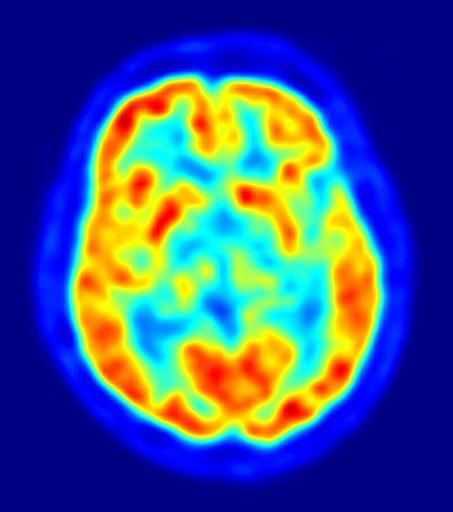
| Keywords: PET-image.jpg This is a transaxial slice of the brain of a 56 year old patient male taken with positron emission tomography PET The injected dose have been 282 MBq of 18F-FDG and the image was generated from a 20 minutes measurement with an ECAT Exact HR+ PET Scanner Red areas show more accumulated tracer substance 18F-FDG and blue areas are regions where low to no tracer have been accumulated Own 2010 Jens Maus http //jens-maus de/ Positron emission tomography | ||||