MAKE A MEME
View Large Image
| View Original: | Nematocyst1.jpg (1347x900) | |||
| Download: | Original | Medium | Small | Thumb |
| Courtesy of: | commons.wikimedia.org | More Like This | ||
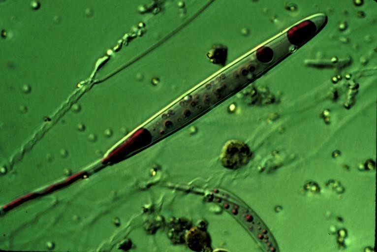
| Keywords: Nematocyst1.jpg Nomarski micrograph of a Ruthenium-red stained nematocyst from Aiptasia pallida the pale anemone The polycationic red dye stains the polyanionic venom proteins found inside the partially discharged nematocyst own work by original uploader ca 1990 David D Brand Dbrand wikipedia en Dbrand Original upload log en wikipedia Nematocyst jpg wikitable - 2007-03-26 14 47 1347×900× 705242 bytes Dbrand <nowiki>Image generated ~1990 by David D Brand Ph D using Nomarski optics </nowiki> - 2007-03-10 23 54 337×225× 24761 bytes Dbrand <nowiki>Nomarski micrograph of a Ruthenium-red stained nematocyst from <i>Aiptasia pallida</i> the pale anemone The red dye stains the polyanionic venom proteins found inside the partially discharged nematocyst </nowiki> - 2007-03-10 23 20 337×225× 24761 bytes Dbrand <nowiki>Nomarski micrograph of a Ruthenium-red stained nematocyst from <i>Aiptasia pallida</i> the pale anemone The reed dye stains the poly-anionic venom proteins found inside the partially discharged nematocyst As the author of this micrograph I hereby re</nowiki> Nematocyst | ||||